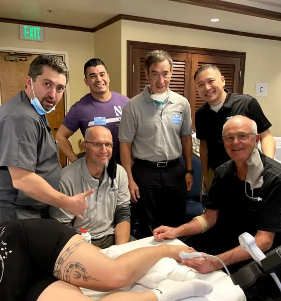
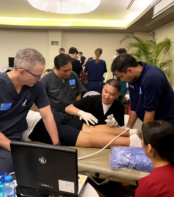
Ultrasound Guided Injection | Cadaver Course Sign Up
FORT WORTH, TX | APRIL 19-20, 2024
The course is designed to provide you with the proper training essential to perform consistent and high quality ultrasound guided injections.
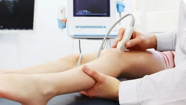
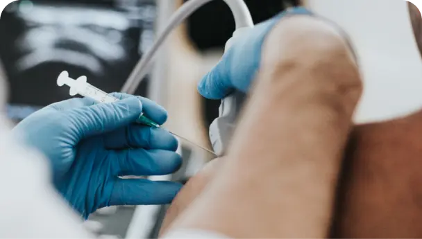
Ultrasound Diagnostic
Course Sign Up
COMING SOON
The course is designed to provide you with the proper training essential to perform consistent and high-quality diagnostic ultrasound exams.
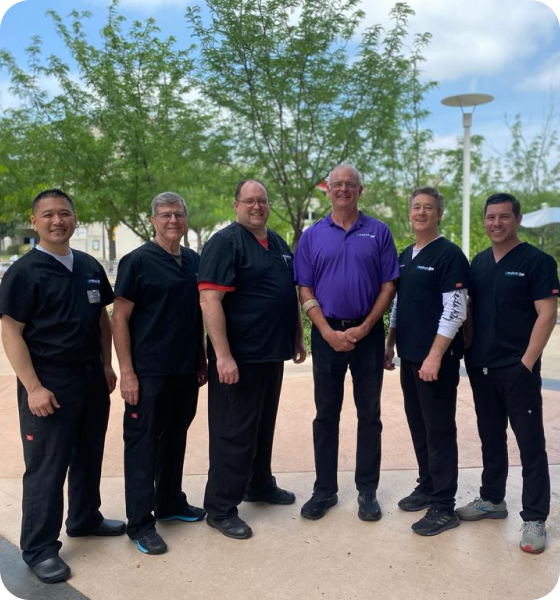
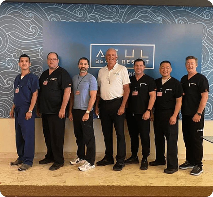
What We Do
MSKUS is dedicated to exceptional training & education in the area of musculoskeletal ultrasound.
Who We Train
Rheumatologists, Orthopedics, Anesthesiologists, Sports Medicine Physicians, Pain Management Physicians, Radiologists, Sonographers.
Accreditation and CME Credit
Working with our experts to complete the training courses and earn CME credits upon successful completion.

Well designed. Great opportunity to practice needle techniques and good references for future study.
Hands-on ultrasound time and instruction at the table was great. 5 of 5 for Dr. Kozar at the table. Great skills and remained patient and amiable with US beginners
Always advance use of ultrasound following these courses. Motivated to review more anatomy (again).
Doing the procedures in a more competent way
Help me to continue to improve skills and provide better patient care.
Great instructors, well organized. Will do it again!
Great instructors, well organized. Will do it again!